
 |  |
Catastrophe always lurks just around the corner in general surgery. The ER pages you, it could be anything. Most of the time it's about a bowel obstruction or appendicitis or a hot gallbladder or a hernia wreaking havoc---issues you deal with every day--- and an almost formulaic mindset is activated. Did you get a CT? NG already in? IV Zosyn please. Go ahead and admit to the regular floor. I'll be there in an hour. And then there are the calls where, 2 minutes into the conversation with the ER doc, you know you are going to have to cancel an elective case or reschedule your afternoon clinic patients to later in the week. Hypotensive, systolics in 70's, rigid abdomen, found down by roommate. We're pumping fluids in. Trying to get a line. WBC 25k, lactate is 13. You know a disaster has just landed on your doorstep and there's nothing to do but drop everything you're doing and go handle it.
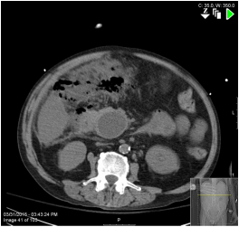
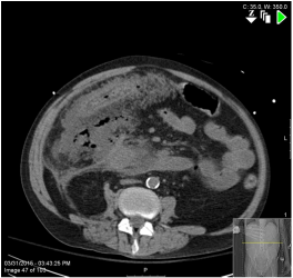
The above images demonstrate a large amount of free air, especially in the lesser sac. A large fluid collection is apparent in the area of the pancreatic head. Mural edema of the right colon is obvious. You call the OR and make arrangements for emergency surgery. You open the poor soul's abdomen and immediately the odor overpowers everyone in the room. You encounter the foul dirty-dishwater fluid and fat saponification pathognomonic of necrotizing pancreatitis. The omentum is plastered to underlying structures encompassing the entire right side of the peritoneal cavity. Gentle finger fracture mobilization of the omentum reveals the catastrophe hiding beneath. Liquefactive necrosis of the hepatic flexure. Complete thrombosis of the vasculature in the mesocolon. You get into the lesser sac and bluntly debride the chunks of pancreatic necrosis, like blackened fragments of dirty curdled milk. The duodenum is mobilized and you find at least two well-demarcated holes in the lateral c-loop. The wall of the duodenum is thinned out, whitish, extremely friable. There's nothing to do here but damage control. The right colon is resected and an end ileostomy is fashioned. Pyloric exclusion to protect the duodeneum. Gastrostomy and jejunostomy tubes for decompression and feeding, respectively. Wash everything out. Put a zillion drains in. Close him up. Get him warm. Fluid and blood product resuscitation. Hope for the best....
The above images demonstrate a large amount of free air, especially in the lesser sac. A large fluid collection is apparent in the area of the pancreatic head. Mural edema of the right colon is obvious. You call the OR and make arrangements for emergency surgery. You open the poor soul's abdomen and immediately the odor overpowers everyone in the room. You encounter the foul dirty-dishwater fluid and fat saponification pathognomonic of necrotizing pancreatitis. The omentum is plastered to underlying structures encompassing the entire right side of the peritoneal cavity. Gentle finger fracture mobilization of the omentum reveals the catastrophe hiding beneath. Liquefactive necrosis of the hepatic flexure. Complete thrombosis of the vasculature in the mesocolon. You get into the lesser sac and bluntly debride the chunks of pancreatic necrosis, like blackened fragments of dirty curdled milk. The duodenum is mobilized and you find at least two well-demarcated holes in the lateral c-loop. The wall of the duodenum is thinned out, whitish, extremely friable. There's nothing to do here but damage control. The right colon is resected and an end ileostomy is fashioned. Pyloric exclusion to protect the duodeneum. Gastrostomy and jejunostomy tubes for decompression and feeding, respectively. Wash everything out. Put a zillion drains in. Close him up. Get him warm. Fluid and blood product resuscitation. Hope for the best....
 RSS Feed